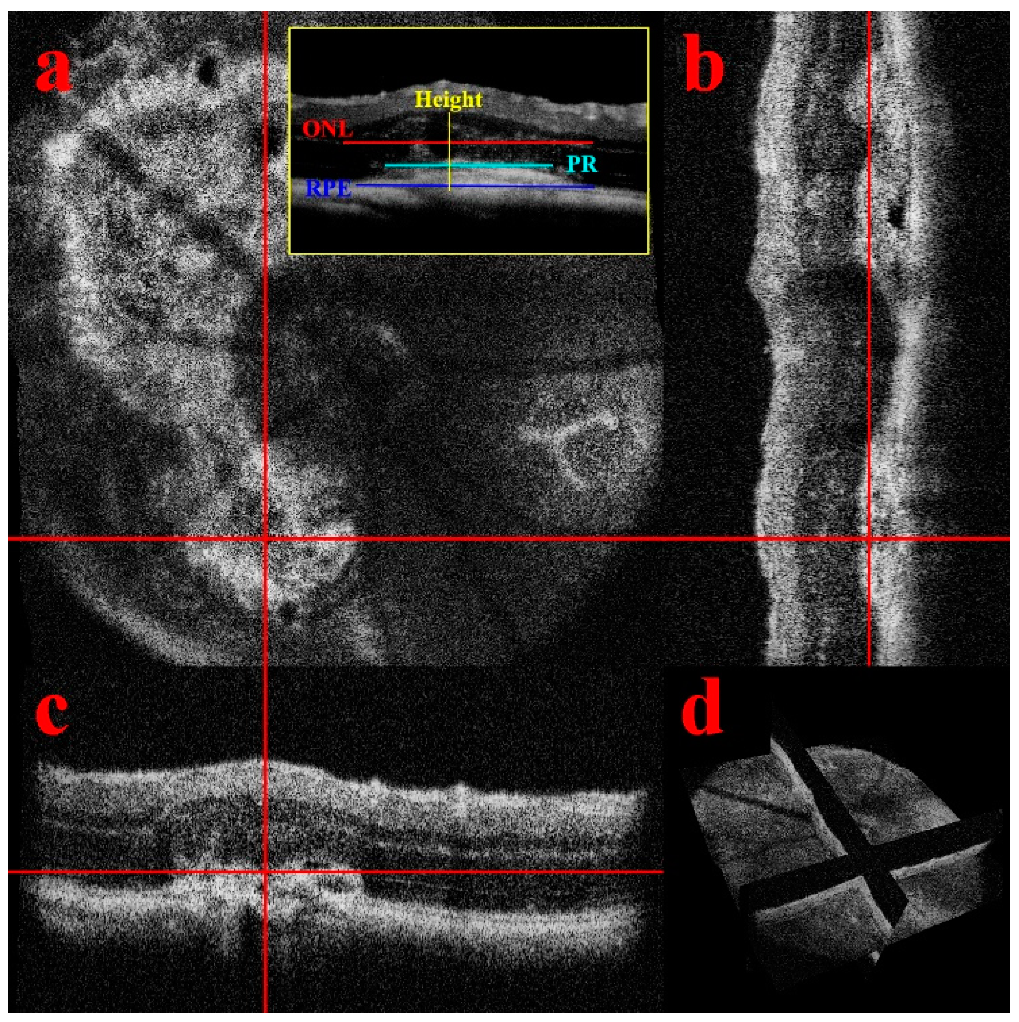
En Face OCT Imaging for the Diagnosis of Outer Retinal Tubulations in Age-Related Macular Degeneration

Postoperative en face optical coherence tomography (OCT) scans of 12... | Download Scientific Diagram
Applied Sciences | Free Full-Text | En-Face Optical Coherence Tomography Angiography for Longitudinal Monitoring of Retinal Injury
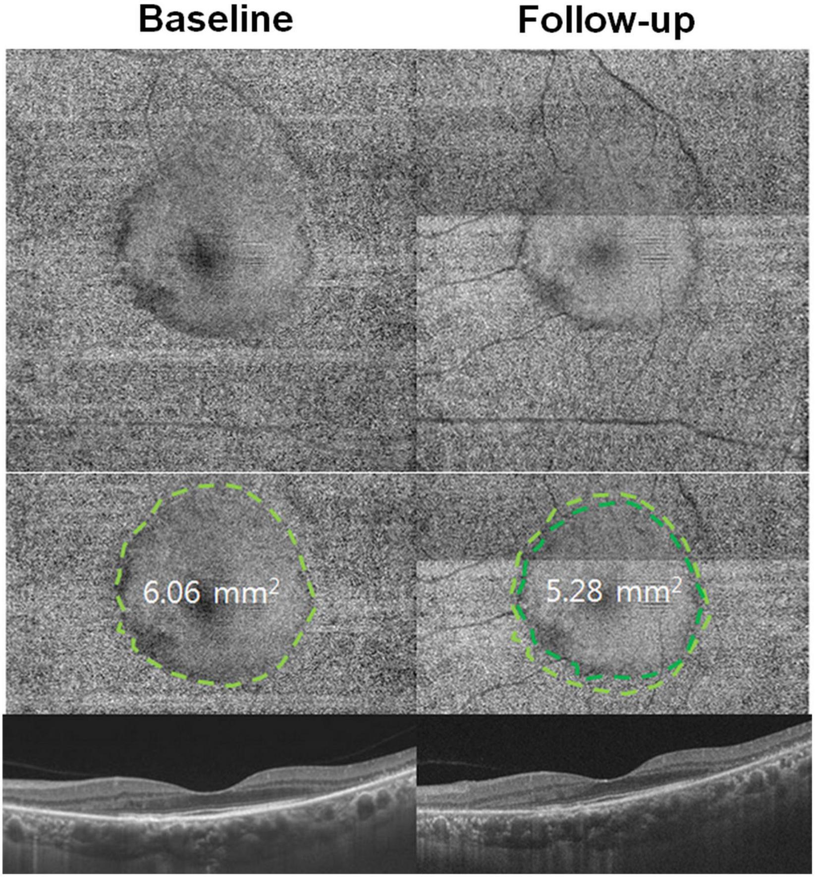
En face image-based classification of diabetic macular edema using swept source optical coherence tomography | Scientific Reports

En Face OCT in Diagnosis of Persistent Subretinal Fluid and Outer Retinal Folds after Rhegmatogenous Retinal Detachment Repair - Ophthalmology Retina

How to acquire the en face optical coherence tomography (OCT) image of... | Download Scientific Diagram
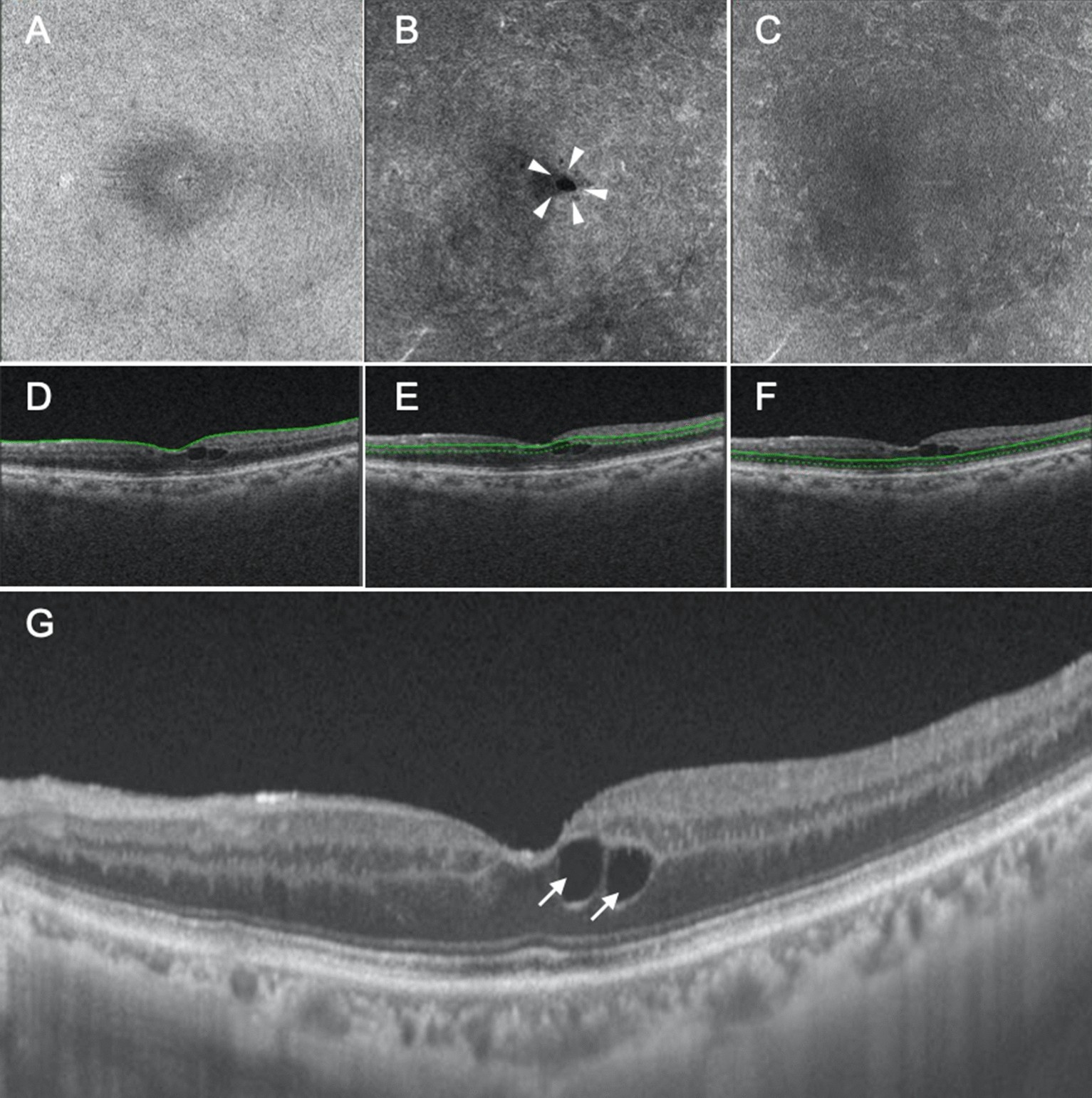
![PDF] En face optical coherence tomography of inner retinal defects after internal limiting membrane peeling for idiopathic macular hole. | Semantic Scholar PDF] En face optical coherence tomography of inner retinal defects after internal limiting membrane peeling for idiopathic macular hole. | Semantic Scholar](https://d3i71xaburhd42.cloudfront.net/2ea6749686dfc9728d5436582047a7d8284a4af1/3-Figure2-1.png)
PDF] En face optical coherence tomography of inner retinal defects after internal limiting membrane peeling for idiopathic macular hole. | Semantic Scholar

En face OCT images generated at two visits in a visually normal subject... | Download Scientific Diagram
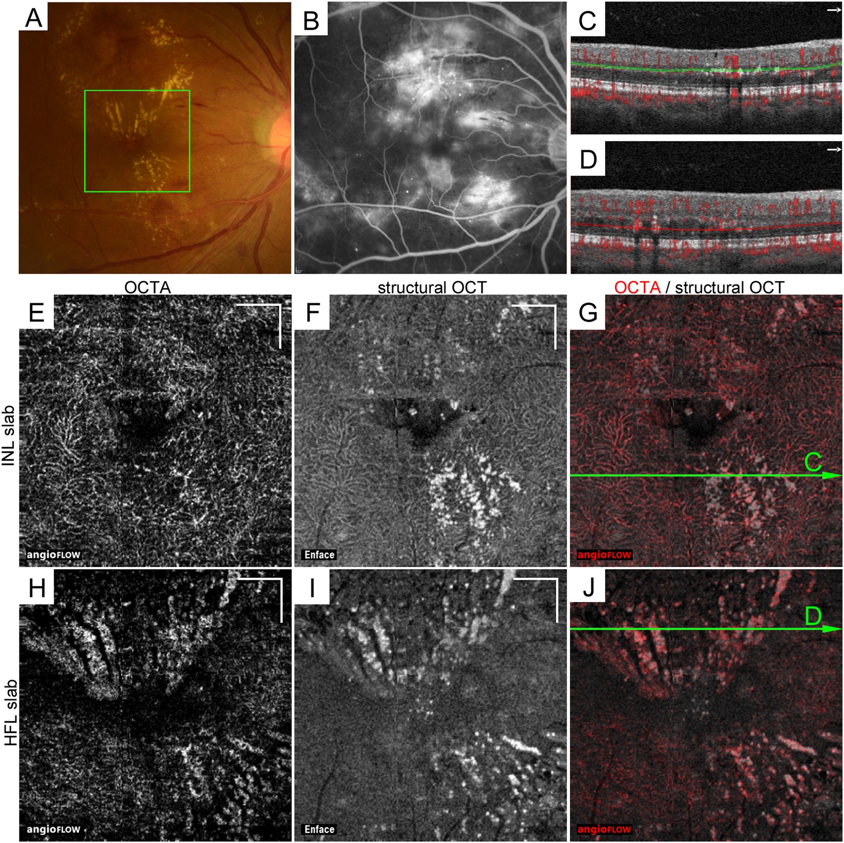
Decorrelation Signal of Diabetic Hyperreflective Foci on Optical Coherence Tomography Angiography | Scientific Reports
![PDF] Intraretinal Correlates of Reticular Pseudodrusen Revealed by Autofluorescence and En Face OCT | Semantic Scholar PDF] Intraretinal Correlates of Reticular Pseudodrusen Revealed by Autofluorescence and En Face OCT | Semantic Scholar](https://d3i71xaburhd42.cloudfront.net/9d329095ddb337f05f47d90c5e84894687872e2a/6-Figure5-1.png)
PDF] Intraretinal Correlates of Reticular Pseudodrusen Revealed by Autofluorescence and En Face OCT | Semantic Scholar
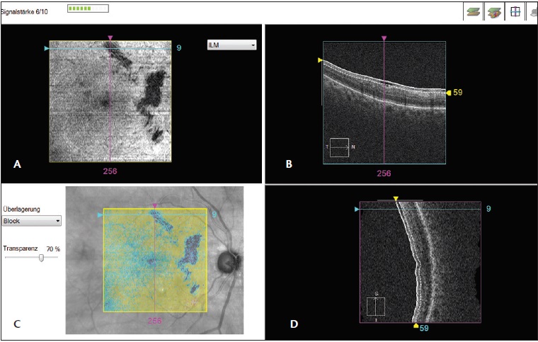
Evaluation of retinal nerve fiber layer defect using wide-field en-face swept-source OCT images by applying the inner limiting membrane flattening | PLOS ONE

Clinical En Face OCT Atlas eBook : Lumbroso, Bruno, Huang, David, Romano, Andre, Rispoli, Marco, Coscas, Gabriel: Amazon.co.uk: Kindle Store
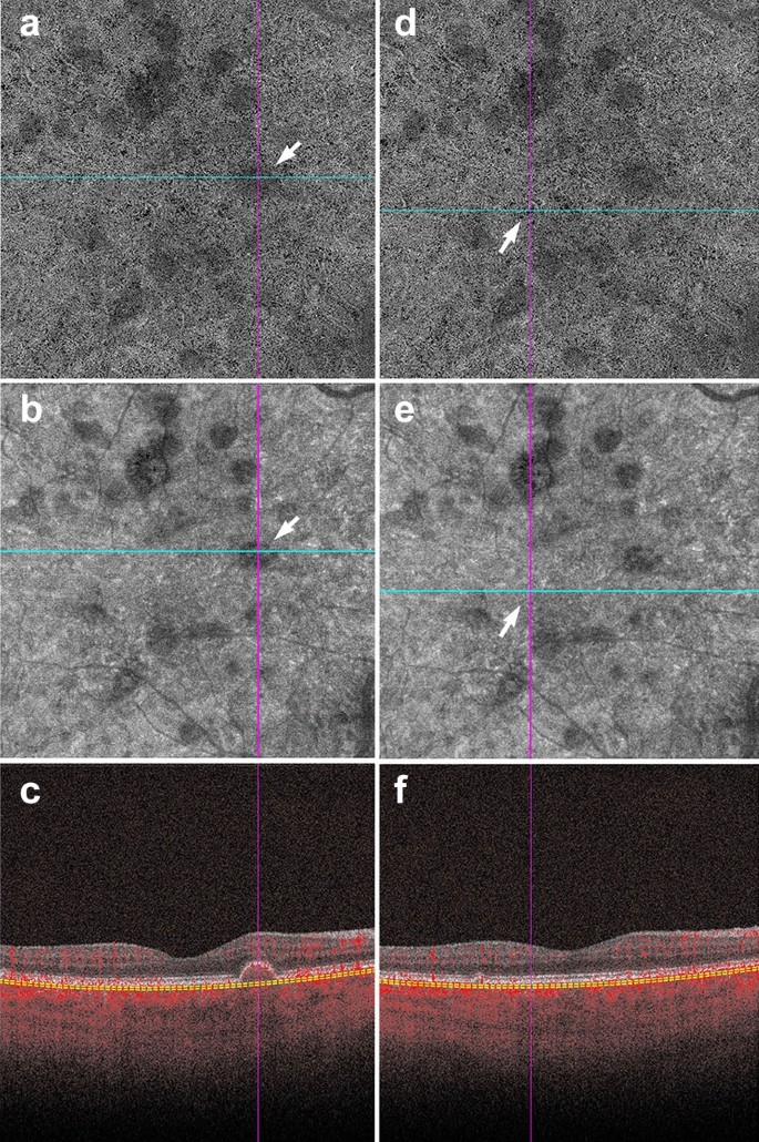
A practical guide to optical coherence tomography angiography interpretation | International Journal of Retina and Vitreous | Full Text
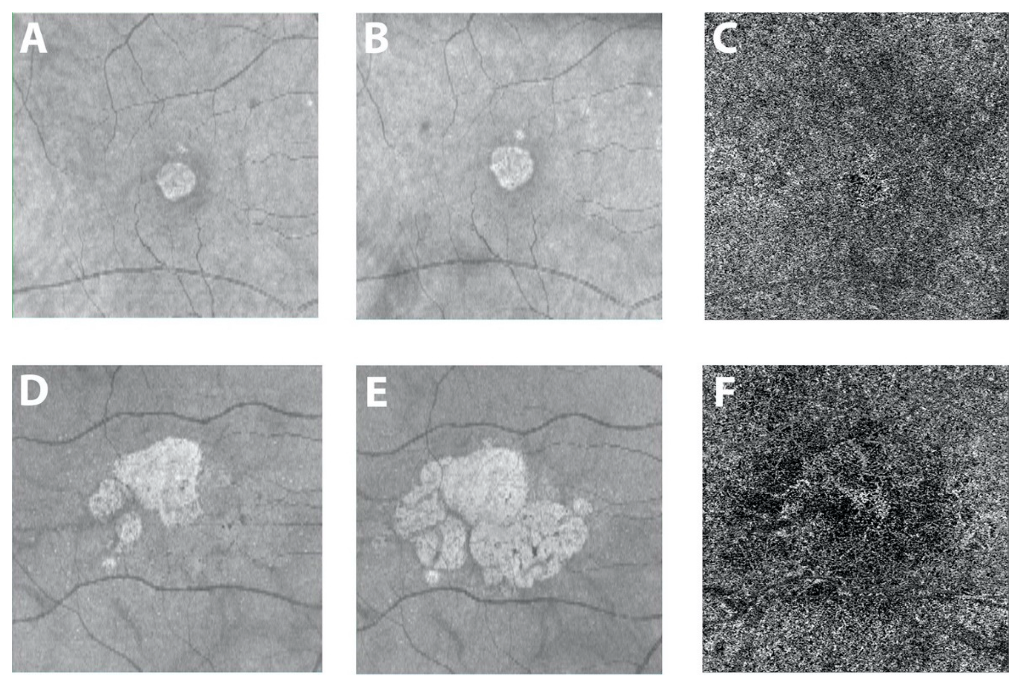
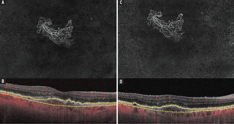
JCM | Free Full-Text | Optical Coherence Tomography Angiography of the Choriocapillaris in Age-Related Macular Degeneration


